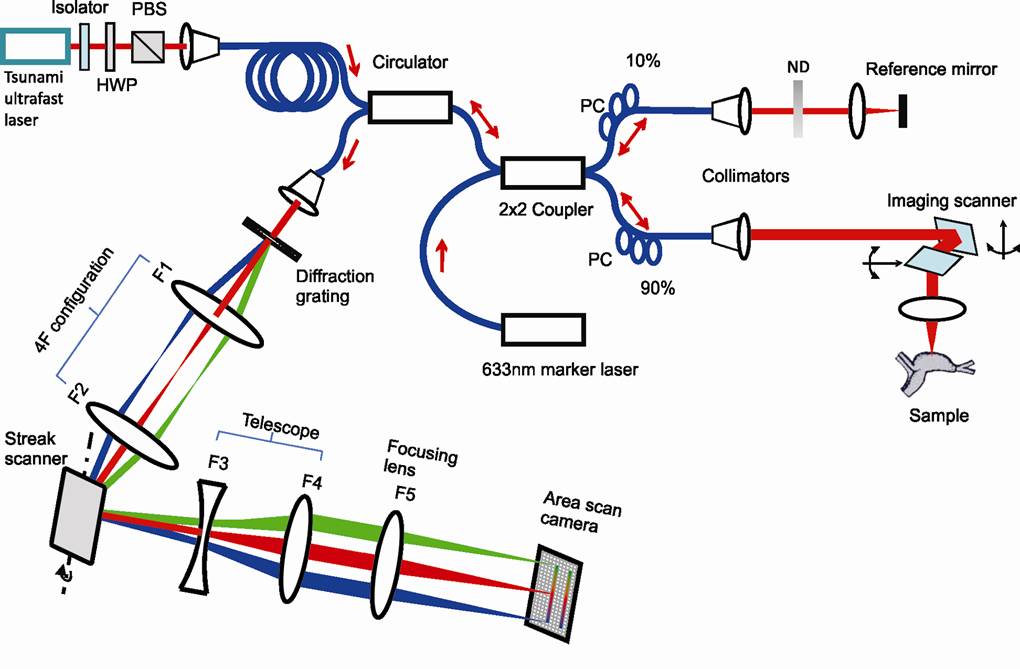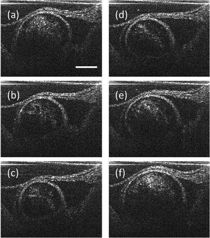
Coherence-based optical imaging technique: In our lab, an ultrahigh-speed Fourier-domain optical coherence tomography (FD-OCT) system is built up that can record the OCT spectrum in streak mode with a high-speed area-scan camera, which allows higher OCT imaging speed than can be achieved with a line-scan camera, shown in Fig. 1. Unlike parallel OCT techniques that also use area-scan cameras, the conventional single-mode fiber based point-scanning mechanism is retained to provide a confocal gate that rejects multiply scattered photons from the sample. When using a 1000 Hz resonant scanner as the streak scanner, 1,016,000 A-scans have been obtained in one second. This method’s effectiveness has been demonstrated by recording in vivo OCT-image sequences of embryonic chick hearts at 1000 frames/s, shown in Fig. 2.
|
Fig. 1 Schematic of the streak-mode FD-OCT. |
Fig. 2 SM-FDOCT image sequence in a single heart stroke of an HH19 chick embryonic heart at 1000 frames/s ( ): (a) begin of the systole ( ); (b) mid-systole; (c) end of the systole; (d) begin of the diastole ( ); (e) mid-diastole; and (f) end of the diastole. Scale bar: 200 μm.
|
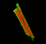
Video of Streak Mode OCT 3D chick heart |
|
|