Medial arterial calcification (MAC) is a common outcome in diabetes and chronic kidney disease (CKD). It occurs as linear mineral deposits along the degraded elastin lamellae and is responsible for increased aortic stiffness and subsequent cardiovascular events. Current treatments for calcification, particularly in CKD, are predominantly focused on regulating the mineral disturbance and other risk factors. Ethylene diamine tetraacetic acid (EDTA), a chelating agent, can resorb mineral deposits, but the systemic delivery of EDTA may cause side effects such as hypocalcemia and bone resorption. We have developed elastin antibody conjugated albumin nanoparticles that target only degraded elastin in vasculature while sparing healthy tissues.
In this study
1, we tested a targeted nanoparticle-based EDTA chelation therapy to reverse CKD-associated MAC. Renal failure was induced in Sprague-Dawley rats by a high adenine diet supplemented by high P and Ca for 28 days that led to MAC. Intravenous delivery of DiR dye-loaded nanoparticles confirmed targeting to vascular degraded elastin and calcification sites within 24 hours. Next, EDTA-loaded albumin nanoparticles conjugated with an anti-elastin antibody were intravenously injected twice a week for two weeks. The targeted nanoparticles delivered EDTA at the site of vascular calcification and reversed mineral deposits without any untoward effects. Systemic EDTA injections or blank nanoparticles were ineffective in reversing MAC. Reversal of calcification seems to be stable as it did not return after the treatment was stopped for an additional four weeks. Targeted EDTA chelation therapy successfully reversed calcification in this adenine rat model of CKD. We consider that targeted nanoparticle (NPs) therapy will provide an attractive option to reverse calcification and has a high potential for clinical translation.

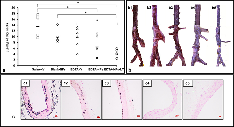
Figure 1: Reversal of aortic calcification by nanoparticle therapy, (n = 6 per group). (a) Calcium quantification in the aortas of all the treatment groups. Saline-IV group showed the maximum amount of calcium per aortic dry weight. Blank-NPs and EDTA-IV groups did not show significantly reduced calcium levels. Both EDTA-NPs* and EDTA-NPs-LT** groups show significant reduction in calcium compared to Saline-IV group. *(One-way ANOVA with Tukey’s HSD) represents statistical significance. Solid line represents mean value. (b) Whole mount aortas stained with alizarin red S to visualize calcium indicated that Saline-IV (b1) and Blank-NPs (b2) group have bright red staining for Ca while slightly reduced red staining in the EDTA-IV group (b3). Alizarin red stain is completely absent in EDTA-NPs (b4) and EDTA-NPs-LT groups (b5). (c) Paraffin-embedded sections of the aortas stained with von Kossa stain (black) revealed Ca deposits in Saline-IV (c1) and Blank-NPs groups (c2) along the degraded elastic lamina and these calcium deposits were slightly reduced in EDTA-IV group (c3). EDTA-NPs (c4) and EDTA-NPs-LT groups (c5) showed no calcium deposits. Scale bar 50
μm.
*EDTA-NPs: anti-elastin antibody-conjugated NPs loaded with EDTA
**EDTA-NPs-LT: anti-elastin antibody-conjugated NPs loaded with EDTA, in which the rats were allowed to survive for 4 weeks after treatment
References
1. Karamched, S. R., Nosoudi, N., Moreland, H. E., Chowdhury, A., & Vyavahare, N. R. (2019). Site-specific chelation therapy with EDTA-loaded albumin nanoparticles reverses arterial calcification in a rat model of chronic kidney disease. Scientific reports, 9(1), 1-11.
