Estrogen Effect on Beak Ligament Structure-Function Relationship in Thumb Basal Joint [Funded by SC INBRE and NIH (NIGMS) COBRE: SC TRIMH]
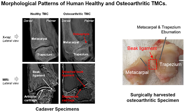
The human thumb basal joint, termed as the trapeziometacarpal (TMC) joint, is the most common site of disabling osteoarthritis (OA) in the upper limb and has a strong predilection for postmenopausal women (10-20:1 women vs. men). Despite extensive pathophysiologic studies in past years, the mechanism of TMC OA with high prevalence in postmenopausal women remains unclear. The laxity of the intracapsular palmar beak ligament, the primary static stabilizer of the TMC joint, has been found to be a pathologic feature of TMC OA. Estrogen, the primary female sex hormone that significantly diminishes after menopause, has been found to significantly affect the cellular biosynthetic and metabolic behaviors in ligamentous tissue (mediated by estrogen receptors).
We hypothesize that primary attritional changes in the beak ligament at the insertions mediated by female sex hormone receptors (i.e., estrogen receptors) affect the structural integrity and mechanical function of the beak ligament. Instability secondary to attrition of the beak ligament is suspected to further change the local mechanical environment inside the basal joint and lead to the progression of OA in postmenopausal females.
The objectives are to examine morphological, mechanoelectrical, and biochemical changes of the beak ligament insertions in relationship to estrogen levels, and further determine the impact of beak ligament laxity/detachment on kinematics and local mechanical environment of the thumb basal joint. The long-term goal of this work is to identify osteoarthritis at early onset and to develop therapeutic strategies to improve functional outcomes.
